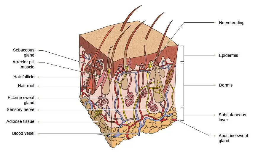
Skin diagram labeled
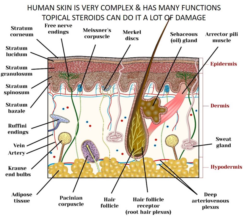
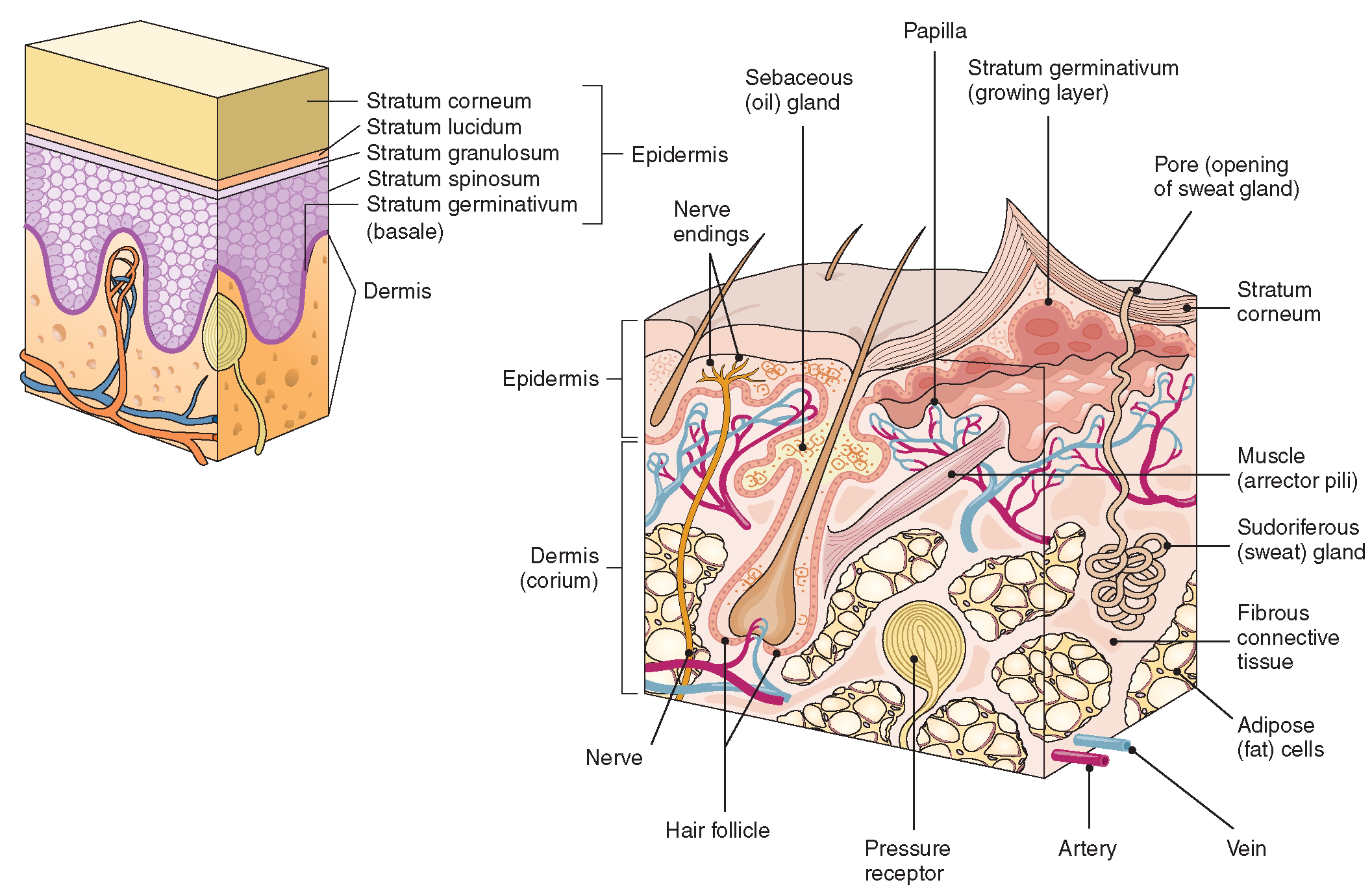
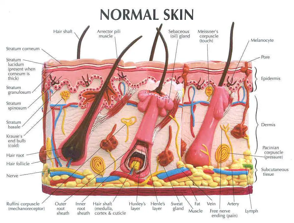
Acne Boils Dandruff Eczema Melanoma It is made up of the following five layers. Stratum Corneum The stratum corneum is the top layer of the epidermis. Its jobs are to: Helps your skin retain moisture Keep unwanted substances out of your body It is made of dead, flattened cells called keratinocytes that are shed approximately every two weeks.
The skin Understanding cancer Macmillan Cancer Support
Read the definitions, then label the skin anatomy diagram below. blood vessels - Tubes that carry blood as it circulates. Arteries bring oxygenated blood from the heart and lungs; veins return oxygen-depleted blood back to the heart and lungs. dermis - (also called the cutis) the layer of the skin just beneath the epidermis.
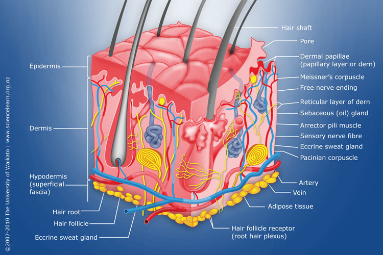
Diagram of human skin structure — Science Learning Hub
The skin is made of multiple layers of cells and tissues, which are held to underlying structures by connective tissue ( Figure 5.1.1 ). The most superficial layer of the skin is the epidermis which is attached to the deeper dermis. Accessory structures, hair, glands, and nails, are found associated with the skin.

Skin Model Labeled Bing Images Skin anatomy, Physiology, Anatomy
This Osmosis High-Yield Note provides an overview of Skin Structures essentials. All Osmosis Notes are clearly laid-out and contain striking images, tables, and diagrams to help visual learners understand complex topics quickly and efficiently. Find more information about Skin Structures: Skin anatomy and physiology. Hair, skin and nails.
Skin topical steroid withdrawal with wheatgrass extract A
Hole's Human Anatomy & Physiology, 9/e. David Shier, Washtenaw Community College. Skin and the Integumentary System. Labeling Exercises. Labeling Exercise 1 Labeling Exercise 2 Labeling Exercise 3 Labeling Exercise 4 Labeling Exercise 5 Labeling Exercise 6: 2002 McGraw-Hill Higher Education
The Integumentary System (Structure and Function) (Nursing) Part 1
Facts about the skin. The skin is the body's largest organ. It covers the entire body. It serves as a protective shield against heat, light, injury, and infection. The skin also: Regulates body temperature. Stores water and fat. Is a sensory organ. Prevents water loss. Prevents entry of bacteria. Acts as a barrier between the organism and its.

Human skin diagram Anatomy and Physiology in 2018 Pinterest Skin
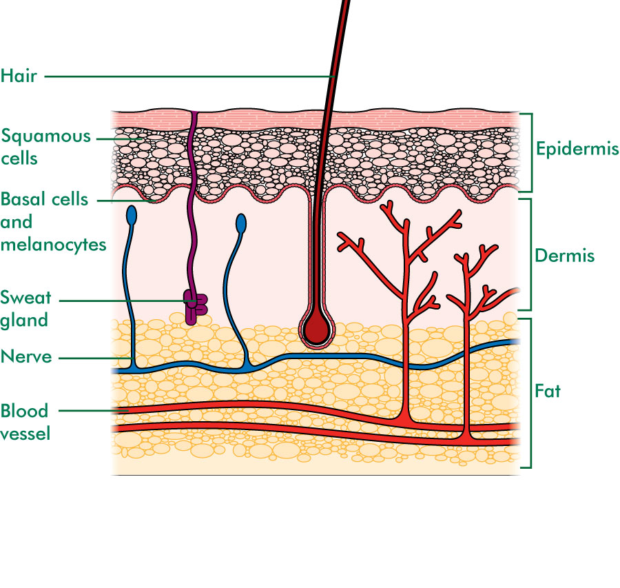
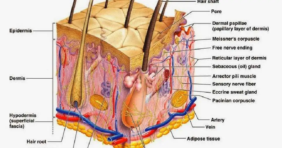
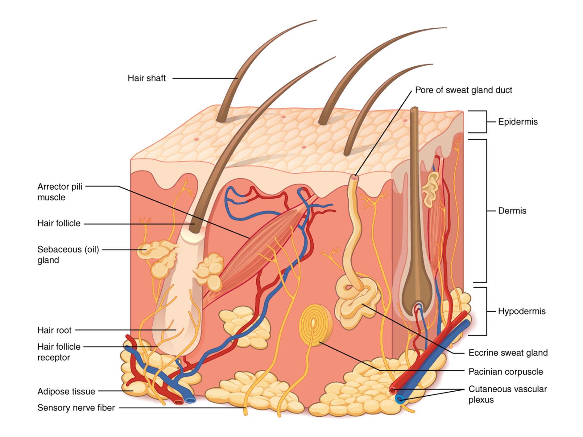
The skin is composed of two main layers: the epidermis, made of closely packed epithelial cells, and the dermis, made of dense, irregular connective tissue that houses blood vessels, hair follicles, sweat glands, and other structures. Beneath the dermis lies the hypodermis, which is composed mainly of loose connective and fatty tissues.

Skin Definition, Structure And Functions Of Skin
Skin. As the body's largest organ, skin protects against germs, regulates body temperature and enables touch (tactile) sensations. The skin's main layers include the epidermis, dermis and hypodermis and is prone to many problems, including skin cancer, acne, wrinkles and rashes. Contents Overview Anatomy Conditions and Disorders Care.
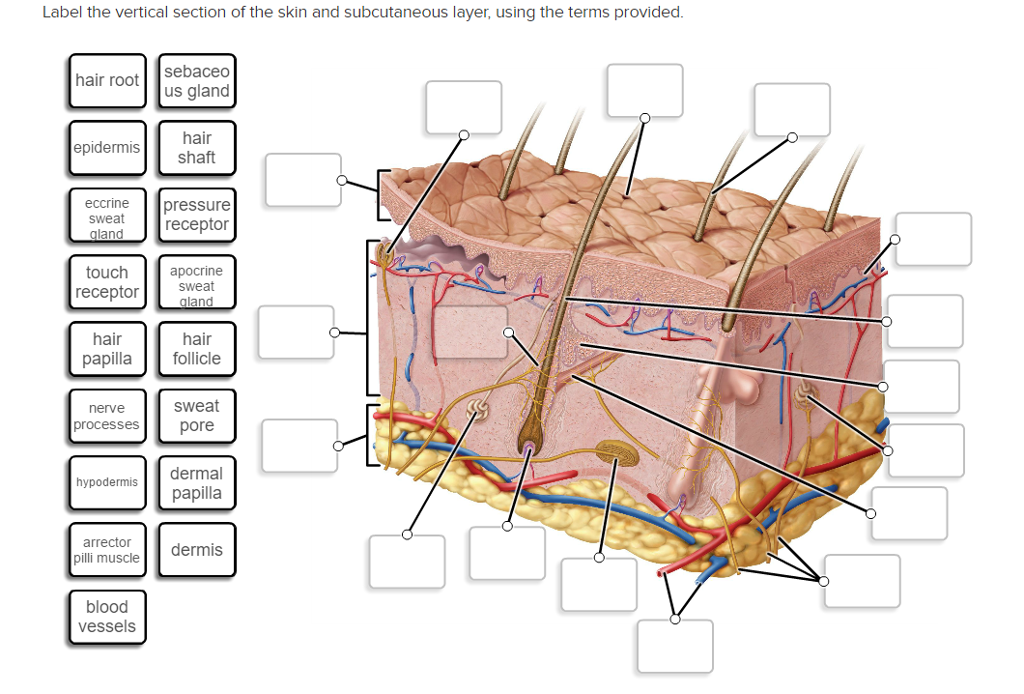
Solved Label the vertical section of the skin and
General histology of the skin. Skin Cutis 1/3 Synonyms: none This article will describe the anatomy and histology of the skin. Undoubtedly, the skin is the largest organ in the human body; literally covering you from head to toe. The organ constitutes almost 8-20% of body mass and has a surface area of approximately 1.6 to 1.8 m2, in an adult.

skin diagram to label Google Search Skin anatomy, Skin structure
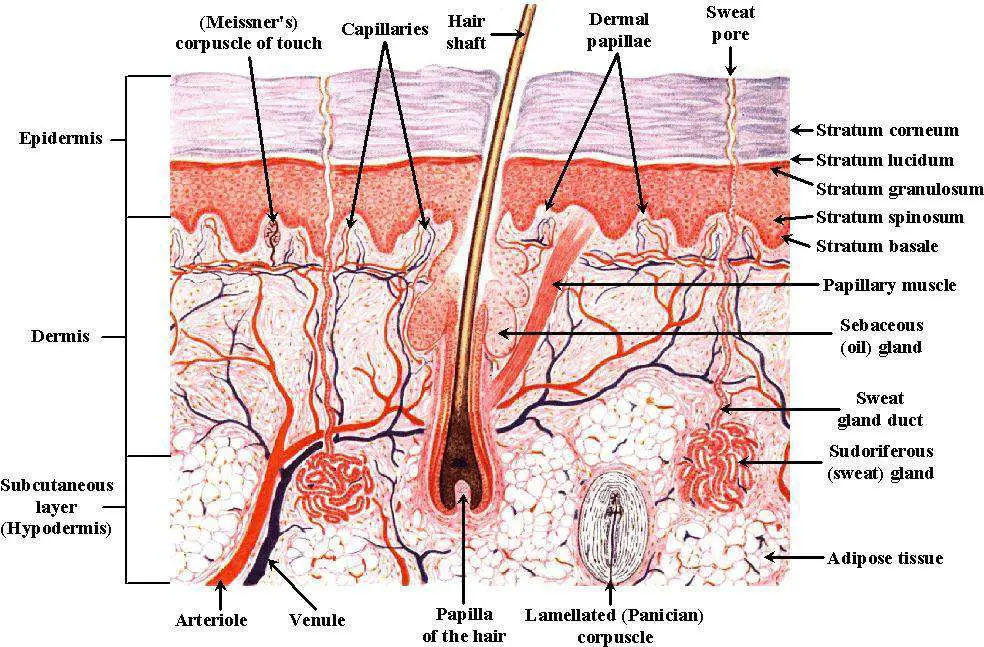
Dermis. Definition. Fibrous and elastic tissue, provides strength and elasticity to the skin and supports the epidermis, home to hair follicles, glands, nerves etc. Location. Term. Papillary Layer. Definition. Upper dermal layer, provides the epidermis with nutrients and regulates body temperature. Location.
Skin diagram to label Labelled diagram
Interactive Link The skin consists of two main layers and a closely associated layer. View this animation to learn more about layers of the skin. What are the basic functions of each of these layers? The Epidermis The epidermis is composed of keratinized, stratified squamous epithelium.
Skin diagram labeled
Epidermis, Dermis, Hypodermis This online quiz is called Skin Labeling. It was created by member marthamae and has 17 questions. Epidermis, Dermis, Hypodermis This online quiz is called Skin Labeling.. Small and Large Intestine Anatomy. Medicine. English. Creator. m_patrick. Quiz Type. Image Quiz. Value. 25 points. Likes. 82. Played. 39,188.

Skin Structure infographic LifeMap Discovery
A hair follicle is a sac-shaped structure in the epidermis in which a hair develops from a group of stem cells. The hair root is the part of a hair that lies below the surface of the skin. It is anchored in the hair follicle. The hair shaft is the visible part of the hair that sticks out of the skin.
Skin diagram labeled
Describe the structure and function of sweat glands and sebaceous glands. Accessory structures of the skin include hair, nails, sweat glands, and sebaceous glands. These structures embryologically originate from the epidermis and can extend down through the dermis into the hypodermis.
Skin diagram labeled
The skin is by far the largest organ of the human body, weighing about 10 pounds (4.5 kg) and measuring about 20 square feet (2 square meters) in surface area. It forms the outer covering for the entire body and protects the internal tissues from the external environment. The skin consists of two distinct layers: the epidermis and the dermis.
POSTECH UNIVERSITY DEVELOPS 3D BIOPRINTING TECHNIQUE THAT GROWS HUMAN
The skin has three layers: Epidermis Dermis Fat layer (also called the subcutaneous layer) Each layer performs specific tasks. Getting Under the Skin The skin has three layers. Beneath the surface of the skin are nerves, nerve endings, glands, hair follicles, and blood vessels.